B21, China Town Mall, Midrand


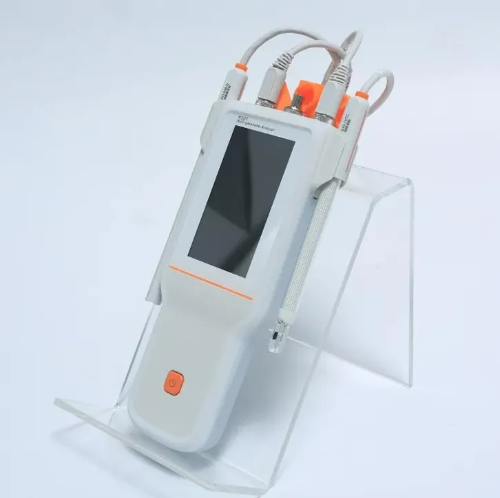
Ophthalmic Diagnosis Portable Handheld Non-mydriatic Digital Fundus Retinal Camera
Asset finance available
Do you want funding for this equipment?
Afrimart can connect you with trusted funding partners who specialise in asset finance for businesses like yours. Compare options and apply in minutes — final approval is subject to provider review.
Check funding options
Secure & no obligation

- Section : Consumer Electronics
- Category : Testing Equipment
- SKU : 1600452933304

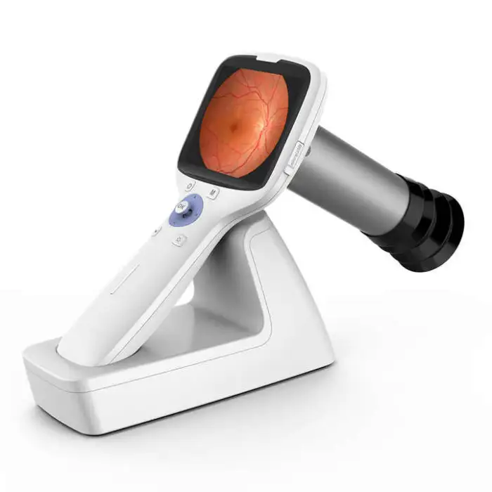
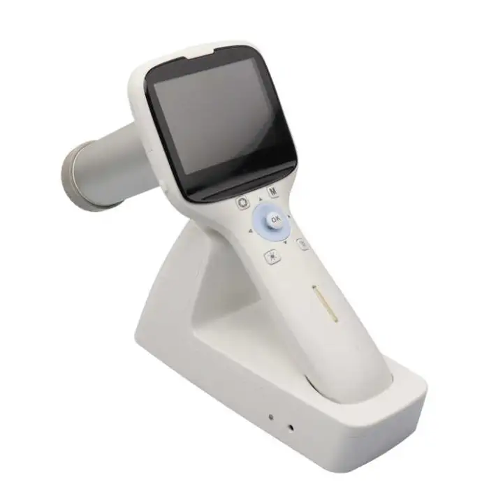
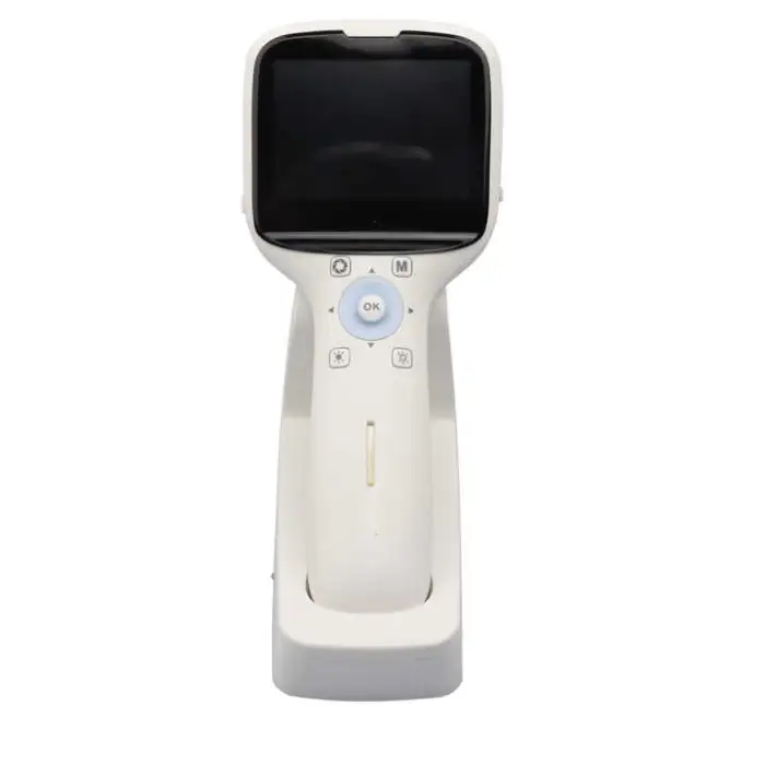
Ophthalmic Diagnosis Portable Handheld Non-mydriatic Digital Fundus Retinal Camera
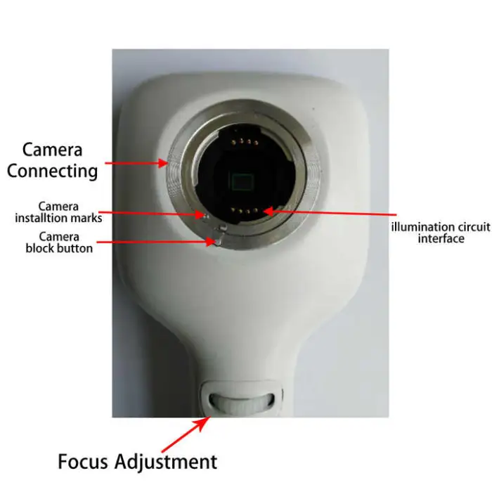
The Ophthalmic Diagnosis Portable Handheld Non-mydriatic Digital Fundus Retinal Camera is a powerful tool for eye care professionals. This compact device allows for easy and quick retinal imaging without the need for pupil dilation. Its user-friendly design and advanced features make it ideal for clinics and hospitals.
Specifications
| Attribute | Details |
|---|---|
| Brand Name | Healicom |
| Model Number | NES-1000 |
| Field of View | 42 degrees |
| Min. Pupil Size | 2.5 mm |
| Screen | 3.5 inch TFT colored screen |
| Image Format | JPEG |
| Image Transfer | Micro USB, WiFi |
| Size | 19 cm * 10 cm * 21 cm |
Key Features
- Portable and easy to use
- No need for pupil dilation
- High-quality retinal imaging
- 3.5 inch TFT colored display for clear viewing
- Supports image transfer via Micro USB and WiFi
Applications
- Routine eye examinations
- Screening for diabetic retinopathy
- Monitoring eye diseases
- Educational purposes in ophthalmology
- Emergency eye care situations
Product Info Gallery
- Shipping Timeframes: All orders are processed within 2-5 business days (excluding weekends and holidays). After your order has been processed, the estimated delivery time is before 07 Sep, 2026, depending on customs, Please note that due to high demand, some items may experience longer shipping times, which will be communicated at order confirmation email.
- Order Processing Time: Please allow 2-5 business days for us to process your order before it is shipped . Orders placed after 16:00 on Fridays, or during weekends and public holidays, will begin processing on the next business day. Processing times may be extended during peak seasons or sales events.
- Manufacturing Time: Some products needs manufacturing time, the manufacturing process will take approximately 10-30 business days depending on the product. This timeframe may vary depending on the complexity of the product and current demand. but this will be communicated with you during order confirmation.
- Returns and Exchanges: We offer a 30-day return policy for most items. If you are not completely satisfied with your purchase, you may return it within 30 days of receipt for a refund or exchange. Items must be unused, in their original packaging, and accompanied by proof of purchase. Return shipping costs are the responsibility of the customer, unless the item was damaged or defective upon arrival.
1. What is the Ophthalmic Diagnosis Portable Handheld Non-mydriatic Digital Fundus Retinal Camera?
It is a compact, handheld device for capturing high-quality retinal (fundus) images without pharmacologic pupil dilation, designed for clinics, hospitals and outreach settings.
2. What are the key specifications of this retinal camera?
Brand: Healicom; Model: NES-1000; Field of view: 42°; Minimum pupil size: 2.5 mm; Display: 3.5-inch TFT color screen; Image format: JPEG; Image transfer: Micro USB and WiFi; Size: 19 × 10 × 21 cm.
3. What does 'non-mydriatic' mean and do I still need to dilate the pupil?
Non-mydriatic means the device can capture retinal images without pharmacologic dilation. However, the camera requires a minimum pupil diameter of 2.5 mm for reliable imaging; dilation may still be needed if the patient’s pupil is smaller or imaging conditions are difficult.
4. What is the field of view (FOV) and why does it matter?
The camera provides a 42-degree field of view, which determines how much of the retina is captured in a single image. A 42° FOV is suitable for standard posterior pole imaging and routine screening.
5. What image formats and transfer options are supported?
Images are saved in JPEG format. Images can be transferred from the device via Micro USB or wirelessly over WiFi for storage, review, or integration into other systems.
6. Does the device have a built-in display for immediate review?
Yes — it includes a 3.5-inch TFT color display for live preview and immediate review of captured images.
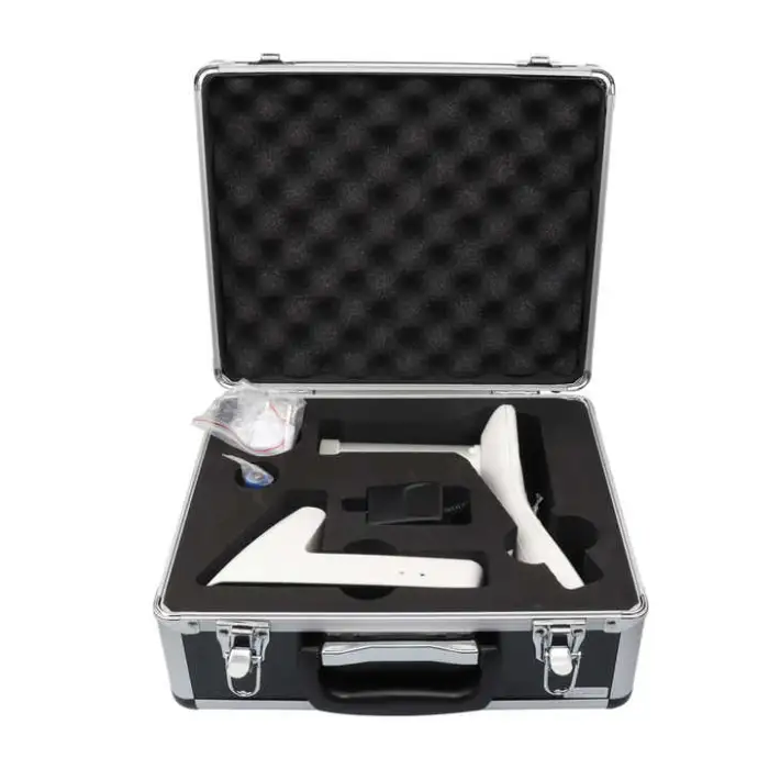
7. Is the camera truly portable and handheld?
Yes. With compact dimensions of 19 × 10 × 21 cm and a handheld design, it is intended for easy transport and use at point-of-care, outreach clinics, and bedside situations.
8. What clinical applications is this camera suitable for?
Common uses include routine eye examinations, diabetic retinopathy screening, monitoring of retinal and optic nerve diseases, ophthalmic education, and emergency eye care triage.
9. What are the recommended infection control or cleaning procedures?
Specific cleaning and disinfection instructions are provided by the manufacturer. Follow those guidelines and local infection-control policies; generally, use appropriate disinfectant wipes that are safe for optical and electronic equipment and avoid liquids entering ports.
10. Is the camera compatible with electronic medical records (EMR) and imaging software?
The camera produces standard JPEG images and supports transfer via Micro USB and WiFi, which enables integration with many systems. For native PACS/EMR integration, confirm supported protocols and software compatibility with the vendor or your IT team.
11. What power or battery options does the device have?
Power and battery specifications are not listed in the provided description. Please consult the manufacturer or product datasheet for details on battery type, runtime, and charging.
12. What training is required to operate the device?
The camera is described as user-friendly and suitable for routine screening, but basic training in fundus imaging technique and patient positioning is recommended. Vendor training or instructional materials can help ensure consistent image quality.
13. Does the product come with warranty and regulatory approvals?
Warranty terms and regulatory clearances (CE, FDA, etc.) are not provided in the description. Contact the manufacturer or authorized distributor for information on warranty, service plans, and applicable regulatory certifications.
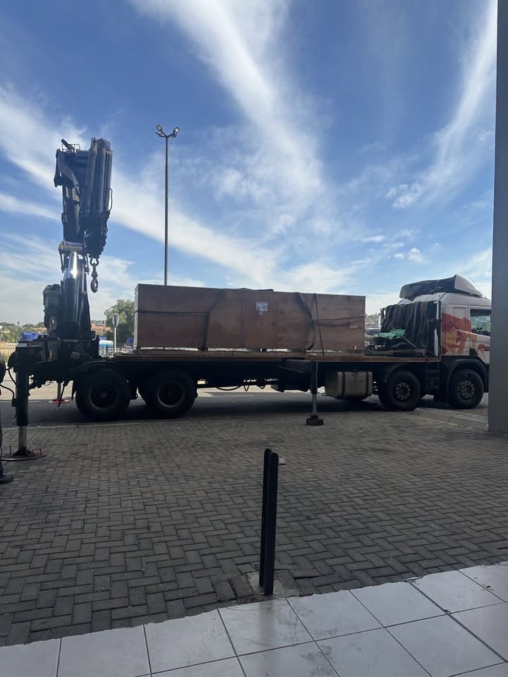
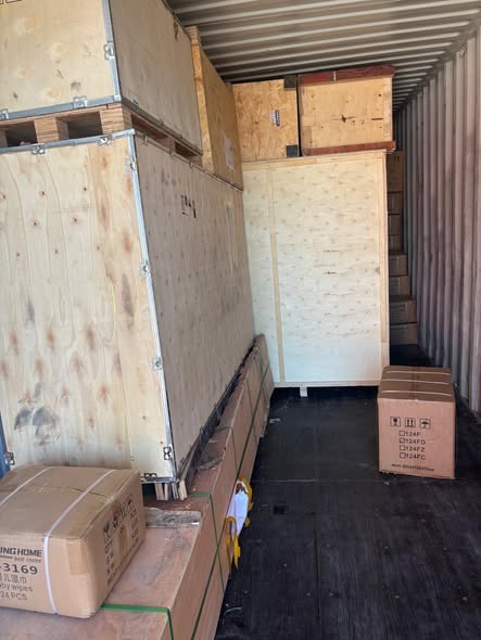
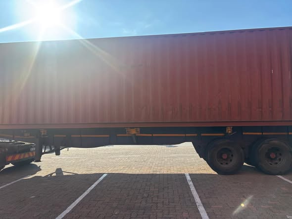
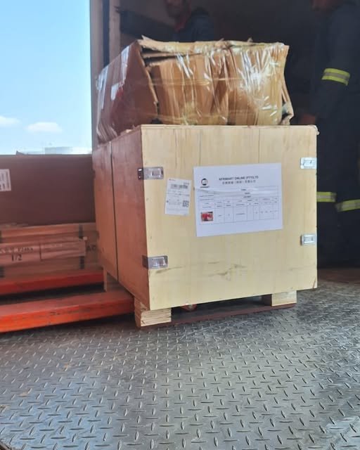
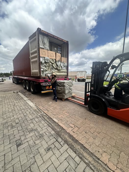
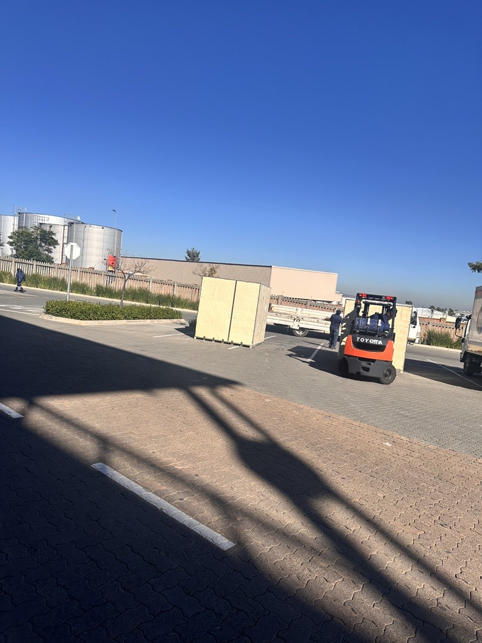
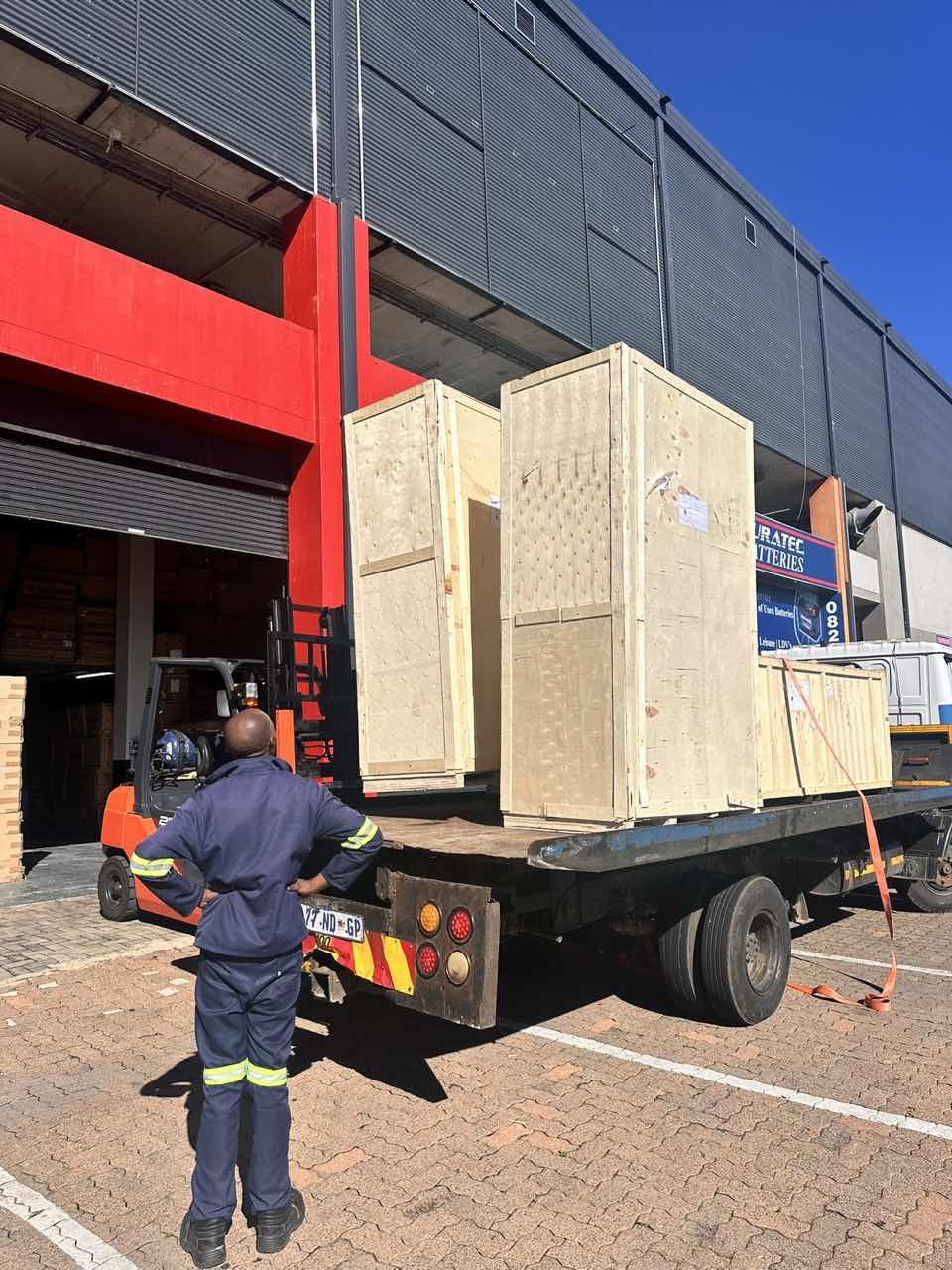
Latest Order Arrivals
Discover our latest orders
12 Heads Embroidery Machine
Toilet Paper Machine Machine

Order Collection
Cross Border Clients
Order cros border countries Collected
Industrial Machine Cllection
Agriculture Processing Machines

Ready for collection

Water Pump Equipment
Packaging Machine and accessories
Fabrics Manufacturing Equipment
Mining Equipments
Food Processing Machine
Batch of Orders
Batch of Orders
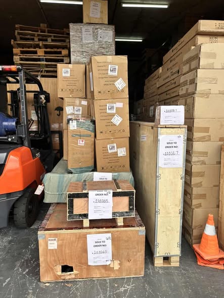
Latest Orders Labelled

wheel alignment machines

new arrivals
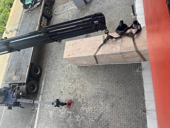
Pre Orders Offloading
Latest Arrivals
Latest Arrivals
Latest Arrivals
Loading
Toilet paper making machine
Toilet paper making machine
Toilet paper Rewinding Machine
latest arrivals
offloading
order success
order collection
order offloading